
Insertional vs Midportion Achilles Tendinopathy: Why the Difference Matters

Insertional and midportion Achilles tendinopathy are not the same condition. Midportion Achilles tendinopathy typically affects the tendon around 2–6 cm above the heel, while insertional Achilles tendinopathy affects the point where the tendon attaches directly to the calcaneus.
That distinction matters because insertional Achilles pain is influenced by both tendon loading and compression against the heel bone, while midportion tendinopathy is predominantly affected by tensile loading and the tendon’s ability to store and release force.
As a result, an exercise or treatment that is appropriate for one type of Achilles tendinopathy may aggravate the other.
At Movement Mechanics in Browns Bay, North Shore Auckland, we assess where the pain is coming from, how the tendon responds to load and whether imaging is needed before deciding whether rehabilitation, radial shockwave therapy, focused shockwave therapy, high-power laser therapy or a combination is appropriate.

What is the difference between insertional and midportion Achilles tendinopathy?
The main difference is the location of the pathology.
Midportion Achilles tendinopathy affects the tendon several centimetres above the heel bone. Insertional Achilles tendinopathy affects the tendon where it attaches directly onto the calcaneus.
The location changes the mechanical environment of the tendon.
Midportion disease is primarily exposed to repeated tensile and energy-storage loading. Insertional disease experiences those same forces but also has to tolerate compression between the Achilles tendon and the posterior heel bone when the ankle moves into dorsiflexion.
That is why the rehabilitation approach should not automatically be identical.

Where does midportion Achilles tendinopathy hurt?
Midportion Achilles tendinopathy usually causes pain approximately 2–6 cm above the heel bone.
Patients often describe:
morning stiffness, tenderness when squeezing the tendon, visible or palpable tendon thickening, pain at the beginning of activity, temporary improvement once warmed up and increased pain after running, walking, jumping or repeated calf loading.
The tendon may feel thick or nodular compared with the other side.
A common description is:
“It is stiff and sore when I first get out of bed, loosens up as I move, then hurts again later.”
This pattern is highly characteristic of Achilles tendinopathy.
Where does insertional Achilles tendinopathy hurt?
Insertional Achilles tendinopathy causes pain directly at the back of the heel where the Achilles attaches to the calcaneus.
Patients commonly notice tenderness at the heel attachment, pain from shoes pressing against the back of the heel, pain with uphill walking or running, discomfort during calf raises and increased symptoms when the ankle moves into deeper dorsiflexion.
Some patients also have associated calcification, enthesophytes, a Haglund-type bony prominence or irritation of nearby bursae.
These findings do not automatically mean the bony or calcific change is the sole cause of pain, but they may influence treatment selection.
How can I tell whether I have insertional or midportion Achilles tendinopathy?
The easiest initial clue is where you can point to the pain with one finger.
Pain directly at the heel attachment suggests insertional Achilles tendinopathy.
Pain several centimetres above the heel is more consistent with midportion tendinopathy.
However, location alone is not enough to make a diagnosis.
A proper assessment should also consider:
how the tendon behaves during calf raises, walking, running and jumping; morning stiffness; local tenderness; tendon thickening; ankle movement; calf strength; whether symptoms are load-related; and whether another condition could be causing posterior heel pain.

Is Achilles tendinopathy the same as Achilles tendonitis?
Not usually. Chronic Achilles pain is better described as tendinopathy rather than tendonitis.
The term “tendonitis” implies inflammation is the main problem.
Persistent Achilles tendinopathy is more complex and is characterised by changes in tendon structure, collagen organisation, cellular activity, vascularity and mechanical capacity.
The tendon has often failed to adapt adequately to the loads being placed on it.
That is why long-term Achilles treatment is not simply about “reducing inflammation”.
The goal is to restore the tendon’s ability to tolerate and produce force.

Why does Achilles tendinopathy become chronic?
Achilles tendinopathy often becomes chronic when tendon loading repeatedly exceeds the tendon’s capacity to recover and adapt.
This may occur after a sudden increase in running volume, hills, speed work, jumping, walking or sport, but symptoms can also develop gradually.
Once chronic changes occur, the tendon may demonstrate collagen disorganisation, altered extracellular matrix, changes in tenocyte activity and altered pain sensitivity.
The result is often a tendon that remains irritable despite periods of rest.

Why is insertional Achilles tendinopathy different mechanically?
Insertional Achilles tendinopathy is affected by compression as well as tensile load.
When the ankle moves forward into dorsiflexion, the lower Achilles tendon can become compressed against the heel bone.
For a healthy tendon this is normally tolerated.
For an irritable insertional tendon, repeated compression can aggravate symptoms.
That is why treatment often initially limits excessive dorsiflexion before gradually restoring tolerance.
Should I stretch insertional Achilles tendinopathy?
Aggressive calf stretching is not always appropriate for insertional Achilles tendinopathy.
Deep calf stretching increases ankle dorsiflexion, which can increase compression at the tendon attachment.
If the insertion is already highly irritable, repeated end-range stretching may worsen symptoms rather than help them.
The better question is not:
“Is stretching good or bad?”
It is:
“Can this particular tendon currently tolerate compression?”
For some patients, stretching can be reintroduced later as irritability improves.
Why can heel drops over a step make insertional Achilles pain worse?
Lowering the heel below the level of a step increases dorsiflexion and compression at the Achilles insertion.
Traditional eccentric heel-drop programmes can be useful in selected Achilles presentations, particularly midportion tendinopathy.
However, in an irritable insertional presentation, repeatedly dropping the heel below neutral may increase compression against the calcaneus and provoke symptoms.
Early insertional rehabilitation often starts with calf loading on level ground before gradually increasing range.
Should I do eccentric exercises for insertional Achilles tendinopathy?
Yes, loading is usually important, but the exercise should be adapted to the insertion.
The problem is not eccentric exercise itself.
The problem is performing it through excessive dorsiflexion when the tendon cannot yet tolerate compression.
A modified calf-strengthening programme may start from the floor or within a restricted range, then progressively increase load and ankle range as symptoms improve.
What exercises are best for midportion Achilles tendinopathy?
Progressive calf strengthening is one of the foundations of treatment for midportion Achilles tendinopathy.
The programme may progress from isometric calf loading to slow isotonic strengthening, heavier calf raises, soleus-specific work, energy-storage exercises, hopping, plyometric loading and eventually running or sport-specific activity.
The exact programme depends on what the patient needs to return to.
A recreational walker does not need the same tendon capacity as a basketball player, sprinter or trail runner.
Can I run with Achilles tendinopathy?
Many patients with Achilles tendinopathy do not need to stop running completely.
Whether running should continue depends on the tendon’s irritability and its response during and after loading.
Important questions include:
Does the pain progressively worsen during the run? Is walking painful afterwards? Is morning stiffness significantly worse the next day? Has calf strength declined? Can you tolerate repeated heel raises? Are hills, speed sessions or long runs driving the flare?
Often the first step is to reduce the highest-load variables rather than eliminate running altogether.
That may mean temporarily reducing hills, intervals, sprinting, long runs or consecutive running days.
How much pain is acceptable when exercising with Achilles tendinopathy?
Some discomfort during rehabilitation can be acceptable, but pain should remain controlled and should not produce a significant deterioration in symptoms the following day.
The tendon’s response over the next 24 hours is often more informative than how it feels during the exercise itself.
A progressive increase in morning stiffness, walking pain or loss of function suggests the current load may be too high.
Why is Achilles pain often worse in the morning?
Morning stiffness is common because tendinopathic tissue often becomes sensitive after a period of inactivity.
The first few steps after waking may feel particularly stiff or painful.
Movement temporarily increases circulation, tissue temperature and neuromuscular activity, which can produce the familiar “warm-up effect”.
However, feeling better once warmed up does not necessarily mean the tendon has fully recovered.

Does Achilles tendinopathy heal with rest?
Rest can reduce symptoms temporarily, but rest alone rarely restores the capacity of a chronically painful Achilles tendon.
The tendon ultimately needs to tolerate load again.
Too much rest can reduce calf and tendon capacity, potentially making return to activity more difficult.
A better strategy is usually relative load modification followed by progressive rehabilitation.
How long does Achilles tendinopathy take to heal?
Achilles tendinopathy usually improves over weeks to months rather than days.
Recovery time depends on:
how long symptoms have been present, insertional versus midportion pathology, tendon structure, strength, loading demands, age, metabolic health, rehabilitation adherence and whether other pathologies are present.
Chronic Achilles tendinopathy that has existed for many months or years may require a longer rehabilitation period.
Importantly, pain improving does not necessarily mean the tendon has regained full capacity.
Can chronic Achilles tendinopathy still heal after years?
Yes. Long-standing Achilles tendinopathy can still improve substantially.
However, chronic cases often require reassessment of:
the original diagnosis, tendon loading, calf strength, ankle mechanics, footwear, training demands and whether the current rehabilitation programme is sufficiently progressive.
Persistent symptoms should not simply lead to repeating the same treatment indefinitely.
Do I need an ultrasound for Achilles tendinopathy?
Not everyone with Achilles pain needs imaging, but ultrasound can be useful when the diagnosis is uncertain or symptoms are persistent.
Ultrasound may identify:
tendon thickening, altered echogenicity, disruption of tendon architecture, vascularity, calcification or other local pathology.
It can also help distinguish Achilles tendinopathy from some alternative causes of posterior heel pain.
At Movement Mechanics, imaging is recommended when it is likely to improve diagnosis, treatment selection or safety rather than being ordered routinely.
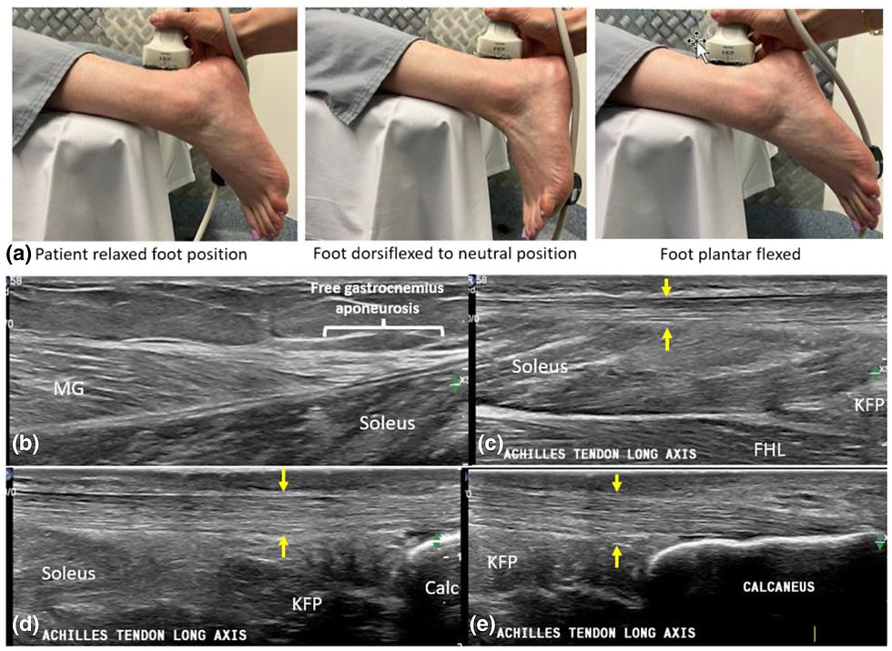
What does Achilles tendinopathy look like on ultrasound?
Typical ultrasound findings can include tendon thickening, heterogeneous structure, hypoechoic regions and altered vascularity.
Insertional disease may additionally show enthesopathic changes or calcification.
However, imaging must always be interpreted alongside symptoms.
Some people have substantial structural change with minimal pain, while others can have significant symptoms with less dramatic imaging findings.

Do I need an MRI for Achilles tendinopathy?
MRI is usually not necessary for straightforward Achilles tendinopathy.
It becomes more useful when there is concern about a partial tear, atypical symptoms, complex insertional pathology, another diagnosis or potential surgical management.
A sudden injury associated with marked weakness or loss of push-off should be assessed for rupture rather than treated as routine tendinopathy.
What else can cause pain at the back of the heel?
Not all posterior heel pain is Achilles tendinopathy.
Other possible causes include retrocalcaneal bursopathy, superficial calcaneal bursitis, partial Achilles tear, plantaris-related pain, posterior ankle impingement, calcaneal stress injury, inflammatory enthesitis, neural irritation and referred pain from the lumbar spine.
That is one reason persistent Achilles pain deserves a proper assessment rather than treatment based only on where it hurts.

Does shockwave therapy help Achilles tendinopathy?
Shockwave therapy can be a useful adjunct for chronic Achilles tendinopathy, particularly when an appropriate rehabilitation programme alone has not produced sufficient improvement.
ESWT applies acoustic mechanical energy to the target tissue.
Research suggests this mechanical stimulus can influence biological processes involved in pain modulation, collagen turnover, vascular signalling, extracellular matrix remodelling and cellular activity.
Shockwave should generally be viewed as an adjunct to rehabilitation rather than a replacement for progressive tendon loading.

How does shockwave therapy work for Achilles tendinopathy?
Shockwave therapy works primarily through mechanotransduction rather than by physically damaging the tendon.
Mechanotransduction describes the process by which mechanical energy is converted into cellular biochemical signalling.
Research shows shockwave exposure can influence pathways associated with:
tenocyte and fibroblast activity, collagen remodelling, VEGF and eNOS signalling, local vascular responses, macrophage behaviour, nociceptive signalling and extracellular matrix regulation.
Some of these mechanisms are supported predominantly by preclinical research, so they should not be confused with direct proof that each mechanism occurs identically in every treated human tendon.
Does shockwave break up scar tissue in the Achilles?
Not in the simplistic sense often described online.
Modern ESWT is better understood as a mechanical stimulus that influences cellular signalling and tissue remodelling.
In calcific pathology, mechanical effects may contribute, but even there the goal is not simply to “smash” calcium or scar tissue.
What is the difference between radial and focused shockwave for Achilles pain?
Radial pressure waves disperse energy over a broader superficial area, while focused ESWT concentrates energy within a defined target at depth.
Radial therapy can be well suited to superficial midportion Achilles tendinopathy and broader treatment of the tendon or calf complex.
Focused ESWT can be particularly useful where the pathology is more focal, deeper, insertional or associated with calcification or the tendon-bone interface.
Neither technology is automatically “better”.
The appropriate source depends on the pathology being treated.

Is focused shockwave better for insertional Achilles tendinopathy?
Focused ESWT can be particularly useful for selected insertional presentations because it allows precise energy delivery to the tendon-bone interface.
This may be relevant where the pathology is highly focal or associated with calcification.
However, not every case of insertional tendinopathy requires focused ESWT.
Clinical examination, imaging where appropriate, tissue irritability and rehabilitation needs should guide the decision.

Is radial shockwave better for midportion Achilles tendinopathy?
Radial shockwave is often practical for superficial midportion Achilles tendinopathy because the target is relatively close to the skin and the treatment field can cover a broader section of tendon.
Radial treatment may also be applied to associated gastrocnemius or soleus tissue where clinically appropriate.
Focused treatment can still be useful in selected midportion cases, particularly when the pathology is focal or radial treatment has not adequately reached the target.

How many shockwave treatments are needed for Achilles tendinopathy?
There is no single treatment number appropriate for every patient.
Clinical protocols commonly involve a short course of treatments separated by approximately one week, with progress assessed over subsequent weeks rather than judging success after the first session.
The appropriate dose depends on the device, acoustic source, target, tissue depth, patient tolerance and clinical response.
How quickly does shockwave therapy work?
Some patients notice pain changes early, but tendon remodelling occurs over a much longer timeframe.
Clinical improvement can continue for several weeks or months after the treatment course.
This is why the success of shockwave therapy should not be judged solely by whether the tendon feels dramatically different immediately after treatment.
Can shockwave make Achilles pain worse before it gets better?
Temporary soreness or an increase in pain can occur after shockwave therapy.
This commonly settles over the following days.
Persistent severe pain, progressive loss of function or new neurological symptoms should be reassessed rather than assumed to be a normal treatment response.
Should shockwave therapy hurt?
Shockwave therapy can be uncomfortable, but more pain does not mean a better treatment.
Energy should be progressed according to tissue tolerance and the treatment objective.
Pain can be used as clinical feedback, but treatment should not become a contest to tolerate the highest possible intensity.
Can laser therapy help Achilles tendinopathy?
High-power laser therapy may be used as an adjunct in selected Achilles presentations, particularly where pain and tissue irritability are limiting rehabilitation.
Photobiomodulation influences cellular signalling through mechanisms different from shockwave therapy, including effects on mitochondrial activity, ATP production, inflammatory signalling, microcirculation and nociceptive sensitivity.
It does not replace progressive rehabilitation.

Can shockwave and laser therapy be used together?
They can be combined when there is a clinical rationale for using two different biological stimuli.
Shockwave provides a mechanical stimulus through mechanotransduction, while laser therapy provides a photobiomodulatory stimulus.
At Movement Mechanics, combination treatment is not used simply because both technologies are available.
The decision depends on the diagnosis, tissue state and treatment goal.
Is a cortisone injection recommended for Achilles tendinopathy?
Corticosteroid injections around the Achilles require considerable caution because of concerns regarding tendon integrity and rupture risk.
For chronic Achilles tendinopathy, treatment usually focuses on progressive rehabilitation and, in appropriate cases, adjunctive therapies such as shockwave rather than routine steroid injection into the tendon.
Any injection decision should be discussed with an appropriately qualified medical practitioner.
When should I worry about Achilles pain?
Seek prompt assessment if Achilles pain follows a sudden injury and is associated with weakness, bruising, a palpable gap or inability to push off normally.
These features raise concern for Achilles rupture.
You should also seek assessment if pain is progressively worsening, walking is becoming difficult, symptoms are unexplained, or rehabilitation is repeatedly failing.
When should I see someone about Achilles tendinopathy?
Assessment is worth considering when Achilles pain has persisted for several weeks, is limiting walking or sport, or repeatedly returns despite rehabilitation.
It is particularly useful when:
you are unsure whether the pain is insertional or midportion, symptoms have persisted for months, previous treatment has failed, imaging has shown significant tendon change, you have had unsuccessful shockwave elsewhere, or you need to return to high-level sport or physically demanding work.
Which treatment is best for Achilles tendinopathy?
There is no single best treatment for every Achilles tendon.
Effective management usually combines accurate diagnosis, appropriate load modification and progressive strengthening.
Shockwave therapy, laser therapy, footwear changes, heel lifts or other interventions can then be added where appropriate.
Treatment should be selected around the pathology rather than around whichever machine or technique happens to be available.
Achilles tendinopathy treatment in Auckland
At Movement Mechanics in Browns Bay, North Shore Auckland, Achilles treatment begins by establishing whether symptoms are coming from the midportion tendon, insertion, surrounding structures or another diagnosis altogether.
Jonathan Hall integrates clinical assessment with:
EMS DolorClast® Radial Shockwave Therapy, EMS DolorClast® Focused Shockwave Therapy, EMS DolorClast® 905 nm High-Power Laser Therapy, osteopathic management, movement assessment, progressive rehabilitation and medical imaging referral where clinically indicated.
Because Movement Mechanics has access to radial shockwave, focused shockwave and high-power laser, treatment can be selected according to the tissue rather than forcing every Achilles presentation through a single modality.
The aim is not simply to reduce symptoms temporarily.
It is to restore the Achilles tendon’s ability to walk, run, jump, climb and tolerate load again.
Movement Matters™
Book an Achilles assessment with Jonathan Hall
If your Achilles has been sore for months despite stretching, strengthening, rest, orthotics or previous treatment, the next step may not be “more of the same”.
The important questions are:
Is the problem insertional or midportion? Is compression contributing? Is the tendon being loaded appropriately? Is imaging required? And would radial shockwave, focused shockwave, laser therapy or rehabilitation alone make the most sense?
Book an assessment with Jonathan Hall at Movement Mechanics, Browns Bay to establish what is driving the problem and build a treatment plan around the actual pathology.
Jonathan Hall M.Ost, BAppSci (Human Biology), PGCertHSc (Acupuncture), GradDipHeal, FIFA Diploma in Football Medicine.
Jonathan is an EMS DolorClast® clinical expert and an ISMST certified practitioner.
Disclaimer: This article is general information, not individual clinical advice. Treatment recommendations require assessment.
© Movement Mechanics Osteopathy Auckland 2024. All rights reserved.


Comments