

Calcific Shoulder Tendinopathy Treatment Auckland
When a scan finally explains the pain, the next question is what to do about it.
There is something strangely reassuring about seeing the problem.
After weeks or months of shoulder pain, an ultrasound or X-ray finally shows a calcium deposit sitting inside the rotator cuff.
For some people, that result makes sense of everything: the night pain, the sharp catch when reaching overhead, the difficulty putting on a jacket, the shoulder that suddenly became almost impossible to lie on.
Then comes the less reassuring part.
Does the calcium need to come out?
The answer is not always.
Calcific shoulder tendinopathy has a natural life cycle. Some deposits disappear without intervention. Others remain embedded within the tendon and continue to cause pain and loss of function. The deposit's appearance, the phase of the condition, and what the shoulder is actually doing all influence what should happen next.
At Movement Mechanics, we use EMS DolorClast® Shockwave Therapy for selected symptomatic calcific rotator cuff deposits, with access to both focused and radial technologies alongside osteopathic assessment and rehabilitation.
For a clearly defined deposit at depth, focused shockwave often gives us the most direct way to deliver treatment to the target.
But the machine is not the first decision.
The deposit is.
Book a Calcific Shoulder Shockwave Assessment
Already Have Imaging? Book an Assessment
Movement Mechanics · Bays Health · Browns Bay · Auckland North Shore · No GP referral required
The Short Answer: Can Shockwave Treat a Calcium Deposit in the Shoulder?
Yes.
Extracorporeal shockwave therapy is one of the better-established non-invasive treatments for persistent calcific rotator cuff tendinopathy.
Clinical trials have demonstrated improvements in pain and shoulder function as well as reductions in the size of calcific deposits. Higher-energy ESWT has generally produced stronger results than lower-energy treatment in chronic calcific presentations. More recent systematic reviews continue to support ESWT compared with sham and also show that other treatments, particularly ultrasound-guided needling or lavage, may be appropriate for some deposits. (Bannuru et al., 2014; Brindisino et al., 2024; Gerdesmeyer et al., 2003).
That does not mean every calcium deposit should receive shockwave.
The shoulder needs to be assessed first.


GDT Certified Clinic
Jonathan is an ISMST Certified Practitioner
No GP referral needed
Appointments available
The Calcium Is Visible. Its Behaviour Is More Important.
Calcific tendinopathy occurs when calcium hydroxyapatite material forms within a rotator cuff tendon.
The supraspinatus is affected most often, although deposits can occur elsewhere in the cuff. Calcific tendinopathy is different from arthritis, and the calcium does not appear because someone has consumed too much calcium in their diet.
The condition is better understood as a cell-mediated tendon process with distinct phases of formation and resorption rather than simple wear and tear. (Uhthoff & Loehr, 1997).
That distinction matters because two shoulder scans can both say “calcific tendinopathy” while describing deposits behaving very differently.
One may be dense, stable and persistent.
Another may already be breaking down.
Treating them identically makes little sense.

Sometimes the Most Painful Shoulder Is the One Already Getting Rid of the Calcium
Calcific shoulder tendinopathy has a recognised biological cycle.
The deposit forms.
It may remain relatively stable.
Eventually, the body can begin actively resorbing it.
During that resorptive phase, inflammatory cells enter the region and the calcium becomes softer and less organised. Material may migrate towards or into the subacromial bursa.
The result can be dramatic.
A shoulder that had been tolerable for months can suddenly become intensely painful at rest and at night. Movement may become extremely difficult despite no significant new injury.
Paradoxically, that severe pain can coincide with the body doing exactly what we ultimately want it to do: removing the deposit.
Current ISMST guidance specifically states that the highly acute calcific shoulder is not an indication for ESWT.
That is why timing matters.
More treatment is not always better treatment.

What Does the Gärtner Classification Tell Us?
When a deposit is visible on X-ray, its appearance can help us understand where it may sit within the calcific cycle.
The commonly used Gärtner classification describes three broad appearances.
The calcium is dense, radiopaque and has clearly defined borders.
This generally represents a more established deposit and is less typical of active spontaneous resorption.


Why Focused Shockwave Enters the Conversation
Most tendon problems do not contain a clearly visible treatment target.
Calcific tendinopathy often does.
The calcium can be seen.
Its location can be measured.
Its depth can be estimated.
And that gives focused shockwave a particular practical advantage.
Focused ESWT concentrates acoustic energy within a defined region beneath the skin. ISMST guidance for calcific tendinopathy specifically describes focused shockwave treatment, with the calcium deposit localised using imaging before treatment.
At Movement Mechanics, our EMS DolorClast® Focused Shock Waves system uses piezoelectric technology to generate and converge acoustic energy within a selected focal region.
For the patient, the physics matters for one simple reason:
We can treat the deposit rather than merely treating the part of the shoulder that hurts.

Does Focused Shockwave “Break Up” the Calcium?
That phrase is useful, but incomplete.
Shockwave is sometimes described as smashing a calcium deposit into pieces.
The biological process appears more complex.
Mechanical stresses generated within and around the calcification may alter the deposit and facilitate fragmentation, while the surrounding biological response supports subsequent cell-mediated resorption.
Clinical studies have shown partial and complete radiographic disappearance of calcifications following ESWT, particularly when adequate energy has been delivered. (Gerdesmeyer et al., 2003; Ioppolo et al., 2012).
What we cannot tell a patient is that every deposit will dissolve.
Some disappear completely.
Some become smaller.
Some change little on imaging despite the shoulder feeling and functioning substantially better.
The X-ray matters.
The person attached to it matters more.

What Does the Evidence Say About Shockwave for Calcific Shoulder Tendinopathy?
The evidence here is considerably stronger than it is for many non-calcific shoulder presentations.
A landmark multicentre randomised controlled trial involving 144 patients with chronic calcific rotator cuff tendinopathy compared high-energy ESWT, low-energy ESWT and sham treatment.
Both shockwave groups improved more than sham, while high-energy treatment produced greater improvement in shoulder function and greater radiographic reduction of the deposits than low-energy treatment. (Gerdesmeyer et al., 2003).
A later systematic review found that high-energy ESWT improved pain, function and calcification resorption in chronic calcific shoulder tendinopathy compared with placebo. (Bannuru et al., 2014).
More recent evidence has added useful nuance.
A 2024 systematic review of 21 randomised trials found ESWT superior to sham for pain and function at 24 weeks and found higher-energy treatment superior to lower-energy ESWT. Importantly, it did not identify a clear difference between focused and radial shockwave for pain, disability or calcification resorption, although the certainty of the evidence was rated very low. (Brindisino et al., 2024).
A 2025 network meta-analysis of 33 randomised trials found comprehensive rehabilitation performed particularly well for function, high-energy ESWT combined with rehabilitation ranked highly for functional improvement, and radial ESWT plus rehabilitation performed strongly for pain. (Yao et al., 2025).
The message is not that one machine solves every calcific shoulder.
It is that appropriate shockwave therapy belongs within a wider rehabilitation strategy.

Focused or Radial Shockwave: Which Is Better for a Calcific Shoulder?
Neither should be described as universally superior.
That distinction is important at Movement Mechanics because we use both.
A randomised study comparing focused, radial and combined ESWT found improvement in pain, movement, function and calcification size across all three groups. (Abo Al-Khair et al., 2021).
The broader 2024 meta-analysis similarly found no clear difference between focused and radial treatment, although certainty was very low.
So why might we still favour focused treatment for some calcific shoulders?
Because the deposit is often a discrete anatomical target located several centimetres beneath the skin.
Focused treatment allows us to select depth and concentrate energy around that target.
Radial treatment may still be appropriate where the deposit and surrounding tissues are readily accessible, or where we also want to treat a broader symptomatic region.
Sometimes both approaches may have a role.
The choice is not:
Which technology is more advanced?
It is:
Which delivery method gives us the most appropriate access to this deposit in this shoulder?
That is a very different question.

The Scan Gives Us a Target. It Does Not Give Us the Whole Diagnosis.
This is where being an osteopathic clinic remains important.
Calcium may explain many of the symptoms.
It doesn't tell us how the shoulder moves now.
A shoulder protected for months can lose range of motion.
The rotator cuff can lose capacity.
The scapula may adopt a different movement strategy.
The thoracic spine may become stiff because the arm is no longer moving normally.
Occasionally, neck or neural symptoms coexist and need to be separated from the shoulder presentation.
We therefore assess the deposit and the person carrying it.
At Movement Mechanics, the osteopathic component may involve examining shoulder range, rotator cuff function, scapular mechanics, and relevant cervical, thoracic, or neurological findings.
Hands-on treatment can be used where a secondary restriction is clinically relevant.
Rehabilitation rebuilds strength and tolerance.
Shockwave is reserved for the pathology it is intended to influence.
The scan provides precision.
Clinical reasoning provides context.

What About High-Power Laser Therapy?
Laser therapy has a different role.
It does not mechanically target a calcium deposit, and we do not describe it as a treatment that dissolves calcium.
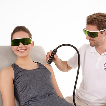
At Movement Mechanics, EMS DolorClast® High-Power Laser Therapy may be considered where the surrounding shoulder is particularly painful or irritable and improving treatment tolerance or movement is the immediate priority.
That becomes especially relevant when the shoulder is too reactive for meaningful mechanical treatment.
Laser and shockwave should therefore not be bundled together automatically.
For some patients, focused shockwave is the main intervention.
For another, radial treatment may be appropriate.
For another, the shoulder first needs to become calmer.
And sometimes the correct treatment is neither.
The benefit of having access to all three EMS technologies is choice, not volume.
What Happens at a Calcific Shoulder Assessment?
If you already have imaging, bring the report.
An X-ray is particularly useful for understanding the deposit's radiographic appearance, while ultrasound can provide additional information about its location, consistency, and the surrounding rotator cuff and bursa.
We look at the calcium itself, but we also ask a different set of questions.
Does the deposit match where you hurt?
Does the shoulder behave like a chronic calcific presentation or an acute resorptive one?
Is movement restricted because of pain, stiffness or something else?
Is the rotator cuff intact?
Is another diagnosis likely to be contributing?
If shockwave is appropriate, imaging helps us determine how to reach the deposit and which EMS technology offers the most sensible treatment strategy.
Treatment can usually begin once we establish suitability.
You are not required to commit to a predetermined course.

How Many Shockwave Treatments Will I Need?
There is no responsible universal answer.
ISMST guidance for calcific shoulder tendinopathy describes focused ESWT delivered in a short treatment course, typically up to five sessions separated by one to two weeks, depending on device technology and clinical circumstances.
Published clinical trials have used different numbers of sessions, energies and treatment intervals.
That variation is exactly why we do not sell “five sessions of shockwave” as though every calcium deposit were identical.
We prescribe treatment according to the presentation.

When Do You Know Whether Shockwave Has Worked?
Not immediately.
Shockwave can influence symptoms relatively early, but calcific resorption and wider tendon adaptation occur over time.
ISMST guidance specifically notes that patients should be counselled that the effect develops over weeks.
That is important when setting expectations.
The shoulder may begin moving more comfortably before an X-ray shows a dramatic structural change.
Conversely, a smaller deposit does not automatically mean somebody has regained the strength and confidence required for normal life.
This is why we reassess function rather than judging success solely by what appears on a scan.

The Outcome Is Not an Empty X-Ray
There is a temptation with calcific tendinopathy to turn treatment into a mission to make the calcium disappear.
That is understandable.
It is also too narrow.
You came because the shoulder was affecting your life.
Perhaps you cannot sleep on it.
Perhaps reaching behind your back has become difficult.
Perhaps you have stopped training.
Perhaps putting on a shirt is painful every morning.
The deposit matters because it may be contributing to those problems.
The reason we treat it is to help restore the shoulder attached to it.
That is why rehabilitation still matters even when shockwave is successful.
The calcium is the target.
Movement is the outcome.
Movement Matters™.

Why Movement Mechanics for Calcific Shoulder Tendinopathy?
Calcific tendinopathy is one of the presentations that influenced our decision to invest in the complete EMS DolorClast® treatment range.
Movement Mechanics provides:
EMS DolorClast® Radial Shockwave Therapy
EMS DolorClast® Focused Shockwave Therapy
EMS DolorClast® High-Power Laser Therapy
within an osteopathic musculoskeletal clinic.
Jonathan Hall is an ACC-registered osteopath, ISMST-certified shockwave practitioner and New Zealand EMS DolorClast® Key Opinion Leader and clinical educator.
That matters less because of the titles themselves than because calcific shoulder treatment is highly dependent on patient selection, imaging, treatment dose and knowing when not to treat.
Access to more technology should not mean receiving more technology.
It should allow a more considered decision.

Calcific Shoulder Shockwave Therapy in Auckland
Movement Mechanics is located inside Bays Health in Browns Bay on Auckland's North Shore.
We see patients from Browns Bay, Torbay, Long Bay, Albany, Mairangi Bay, Milford, Takapuna and across greater Auckland who already have imaging confirming calcific rotator cuff tendinopathy or who have been advised that calcium may be contributing to their shoulder pain.
You do not need a GP referral.
Referrals from GPs, physiotherapists, surgeons and other health professionals are also welcome.
If your scan already shows a calcium deposit, bring the report with you.
We can start by answering the question that matters most:
Is this deposit suitable for shockwave therapy?
Book a Calcific Shoulder Shockwave Assessment

Your Scan Has Shown Us Where to Look
A calcium deposit gives us something unusually concrete in musculoskeletal medicine.
We can see it.
We can measure it.
And in the right circumstances, we can direct treatment towards it.
But the quality of the decision still matters more than the clarity of the X-ray.
A deposit already undergoing resorption may need time and symptom management.
A chronic, well-defined deposit may be an excellent candidate for shockwave.
A soft deposit may make lavage worth considering.
And a shoulder with another significant pathology may need an entirely different pathway.
The purpose of an assessment is to know the difference.
Movement Mechanics. Movement Matters™.
Bring your X-ray, ultrasound or MRI report if you already have one.
Written and clinically reviewed by Jonathan Hall
M.Ost, BAppSci (Human Biology), PGCertHSc, GradDipHeal
ISMST Certified Shockwave Therapy Practitioner
Founder & Clinical Director, Movement Mechanics
New Zealand EMS DolorClast® Key Opinion Leader
Last clinically reviewed: September 2026
Reference
-
Abo Al-Khair, M. A., El Khouly, R. M., Khodair, S. A., Al Sattar Elsergany, M. A., Hussein, M. I., & Mowafy, M. E. E. (2021). Focused, radial and combined shock wave therapy in treatment of calcific shoulder tendinopathy. The Physician and Sportsmedicine, 49(4), 480–487. https://doi.org/10.1080/00913847.2020.1856633
-
Bannuru, R. R., Flavin, N. E., Vaysbrot, E., Harvey, W., & McAlindon, T. E. (2014). High-energy extracorporeal shock-wave therapy for treating chronic calcific tendinitis of the shoulder: A systematic review. Annals of Internal Medicine, 160(8), 542–549. https://doi.org/10.7326/M13-1982
-
Brindisino, F., Marruganti, S., Lorusso, D., Cavaggion, C., & Ristori, D. (2024). The effectiveness of extracorporeal shock wave therapy for rotator cuff calcific tendinopathy: A systematic review with meta-analysis. Physiotherapy Research International, 29(3), e2106. https://doi.org/10.1002/pri.2106
-
EMS Electro Medical Systems. (n.d.). Calcifying tendinitis of the shoulder. EMS Pain Therapy. https://www.ems-dolorclast.com/indications/calcifying-tendinitis-shoulder
-
Forogh, B., Karami, A., & Bagherzadeh Cham, M. (2024). Effect of extracorporeal shock wave therapy and ultrasound-guided percutaneous lavage in reducing the pain of rotator cuff calcific tendinopathy: An updated systematic review and meta-analysis. Journal of Orthopaedics, 56, 151–160. https://doi.org/10.1016/j.jor.2024.05.023
-
Gerdesmeyer, L., Wagenpfeil, S., Haake, M., Maier, M., Loew, M., Wörtler, K., Lampe, R., Seil, R., Handle, G., Gassel, S., & Rompe, J. D. (2003). Extracorporeal shock wave therapy for the treatment of chronic calcifying tendonitis of the rotator cuff: A randomized controlled trial. JAMA, 290(19), 2573–2580. https://doi.org/10.1001/jama.290.19.2573
-
International Society for Medical Shockwave Treatment. (2023). ESWT guidelines: English version (3rd ed.). https://shockwavetherapy.org/wp-content/uploads/2024/01/ISMST-Guidelines-for-ESWT-_-engl-20240103.pdf
-
Ioppolo, F., Tattoli, M., Di Sante, L., Attanasi, C., Venditto, T., Servidio, M., Cacchio, A., & Santilli, V. (2012). Extracorporeal shock-wave therapy for supraspinatus calcifying tendinitis: A randomized clinical trial comparing two different energy levels. Physical Therapy, 92(11), 1376–1385. https://doi.org/10.2522/ptj.20110252
-
Uhthoff, H. K., & Loehr, J. W. (1997). Calcific tendinopathy of the rotator cuff: Pathogenesis, diagnosis, and management. Journal of the American Academy of Orthopaedic Surgeons, 5(4), 183–191. https://doi.org/10.5435/00124635-199707000-00001
-
Yao, Y., Yang, G., Jiang, S., Ji, B., Jin, H., Tang, P., Li, H., Lu, B., & Li, Y. (2025). Treatments for rotator cuff calcific tendinitis: A systematic review and network meta-analysis of randomized-controlled trials. EFORT Open Reviews, 10(7), 520–533. https://doi.org/10.1530/EOR-2024-0078
Disclaimer: This content is for educational purposes and does not constitute medical advice. Individual patient suitability for perioperative shockwave and laser therapy should be assessed by a qualified healthcare practitioner in direct collaboration with the treating surgical team. Contraindications to ESWT and photobiomodulation must be screened for prior to treatment.